Can you get this one ?

Dunga Sai Kumar MBBS, DNB (General Medicine), DM (Clinical Immunology and Rheumatology)
<br
Consultant Rheumatologist, Apollo Hospitals, Arilova, Visakhapatnam</br
Case 1 details
A 24-year-old male presented with right hip pain and inflammatory backache for 6 months. Upon examination, he had swollen and tender bilateral sternoclavicular joints. He also had Pustules involving the scalp (B) and also had acneform eruption involving the upper chest (C) and back region. X-ray showed bilateral sacroiliitis. Tc99m-MDP three-phase bone scan showed increased tracer concentration in bilateral sternoclavicular joints, bilateral 1st and costochondral joints, bilateral sacroiliac joints, and hip joints (C, D). His inflammatory markers were elevated and HLA b27 was Negative. His symptoms improved upon initiating NSAIDs and DMARDs along with low-dose steroids.

1. What is the diagnosis?
2. What is the name of the sign in the Bone scan?
Answer Key:
- SAPHO syndrome (Synovitis, Acne, Pustulosis, Hyperostosis, Osteitis)
- Bull’s Head sign
Suggested reading
- Liu S, Tang M, Cao Y, Li C. Synovitis, acne, pustulosis, hyperostosis, and osteitis syndrome: review and update. Ther Adv Musculoskelet Dis. 2020;12:1759720X20912865. Published 2020 May 12. doi:10.1177/1759720X20912865
- İlgen U, Turan S, Emmungil H. Bull’s Head Sign in a Patient with SAPHO Syndrome. Balkan Med J. 2019;36(2):139-140. doi:10.4274/balkanmedj.galenos.2018.2018.1630
Case 2 –The enigma of unyielding mucosal infection.
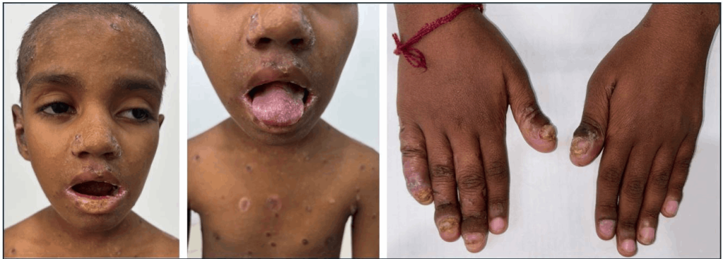
A 4.5-year-old male child born out of a nonconsanguineous marriage presented with a history of recurrent oral thrush, lip erosions, and nail dystrophy starting at 2 months of age, accompanied by a failure to thrive. At 2 months, he developed white patches in the oral cavity (oral thrush), along with erythema and cracking of the lips. Despite repeated antifungal treatments (nystatin, fluconazole), the oral candidiasis recurred. By 6 months of age, progressive nail dystrophy, including thickening, discoloration, and brittleness of the nails, affects both hands and feet. There was no improvement either with multiple courses of topical and oral antifungals or systemic antibiotics for presumed bacterial infections.

1. What is the diagnosis
2. Which cytokine is implicated in this disease pathogenesis
Answer Key:
- Chronic Mucocutaneous candidiasis (The child’s genetic workup showed positive STAT1 heterozygous pathogenic mutation with autosomal dominant inheritance)
- IL-17
Suggested reading
- Shamriz O, Tal Y, Talmon A, Nahum A. Chronic Mucocutaneous Candidiasis in Early Life: Insights Into Immune Mechanisms and Novel Targeted Therapies. Front Immunol. 2020;11:593289. Published 2020 Oct 16. doi:10.3389/fimmu.2020.593289

