Nidhish Chandra MD, DNB (Gold Medalist) Senior Resident, Department of Clinical Immunology & Rheumatology, King George Medical University, Lucknow
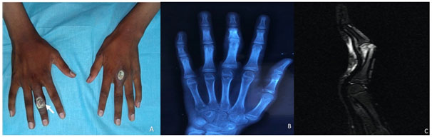
A 17-year-old boy presented to the rheumatology unit with a one-year history of bilateral symmetrical proximal muscle weakness and erythematous papules over the left third metacarpophalangeal and right third proximal interphalangeal joints with subsequent ulceration. He also reported purulent discharge with severe pain and restriction of movement of the right digit (Panel A). The discharge resolved spontaneously before his presentation to our clinic. The history of severe pain and discharge prompted further evaluation with a radiographand MRI of the right hand.
Juvenile dermatomyositis-likely anti-TIF1y/NXP2 or MDA-5 due to ulcerated Gottron’s papule.
Osteomyelitis with cortical irregularity of the right third middle phalanx on radiograph and bone marrow oedema with surrounding soft tissue oedema on MRI